



MBBS (Singapore), MRCS (Edinburgh), MMed (Ortho), FRCS (Ortho), FAMS (Ortho)
Your MRI says you have mild or moderate spinal stenosis, but what does that actually mean for you? The terminology can be confusing, especially when the severity described on your scan does not seem to match what you are experiencing.
An MRI can identify changes within the spine, but the wording of the report does not tell the full story. Understanding how these findings are interpreted alongside your symptoms can give you a clearer picture of what the result means for your spinal health.
What Can an MRI Show About Spinal Stenosis?
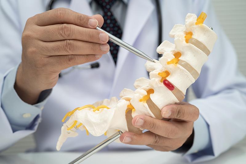
An MRI provides detailed images of the spinal canal, discs, nerves, spinal cord and surrounding soft tissues. It can help identify where stenosis is located and which structures may be affected.
The location also provides important context. Cervical spinal stenosis affects the neck, while lumbar spinal stenosis occurs in the lower back.
What Do Mild and Moderate Spinal Stenosis Mean?
Terms such as mild, moderate and severe may be used in an MRI report to describe the extent of narrowing seen on imaging. The exact criteria used to describe severity can vary depending on the location and type of stenosis. These terms describe the imaging findings rather than directly measuring the severity of your symptoms.
Mild Spinal Stenosis
Mild stenosis generally means that a relatively small degree of narrowing is visible on the MRI. The significance of the finding depends on where the narrowing occurs and whether it affects nearby nerves or the spinal cord. Some people with mild stenosis have symptoms, while others have no related symptoms.
Moderate Spinal Stenosis
Moderate stenosis indicates a greater degree of narrowing on the MRI. Depending on its location, there may be less space around a nerve or the spinal cord, and there may be evidence of contact or compression. However, a moderate MRI finding does not automatically mean that you will have moderate symptoms or require more intensive treatment.
Why Does MRI Severity Not Always Match Your Symptoms?
MRI findings and symptom severity do not always correspond closely. Two people with similar-looking scans can have very different experiences.
Several factors help explain this difference:
- Location of narrowing: Stenosis only becomes relevant to a particular symptom if it affects structures associated with that area of the body
- Degree of nerve involvement: The amount of visible narrowing does not always reflect how irritated or affected a nerve is
- Individual anatomy: The amount of space naturally available within the spinal canal differs between individuals
- Other spinal changes: Disc problems, arthritis or inflammation may contribute to symptoms alongside stenosis
- Activity and position: Some symptoms become more noticeable during particular movements or activities rather than remaining constant
For this reason, your MRI grade should not be interpreted on its own.
How Are Your MRI Findings Interpreted?
Your doctor will compare the MRI findings with your symptoms, medical history and physical examination. This helps determine whether the narrowing shown on the scan is actually responsible for what you are experiencing.
Depending on the location of the stenosis, your assessment may include muscle strength, sensation, reflexes, hand coordination, balance and walking. Your doctor will also consider where you experience pain, numbness or tingling and whether this corresponds with the nerves affected on the MRI. This helps determine whether an MRI finding is related to your symptoms or simply an age-related change that is not causing your current problem.
How MRI Findings Influence Treatment Decisions
While MRI findings provide important information about spinal narrowing, the grade alone does not determine your treatment. Your symptoms, neurological function and examination findings are also considered.
Mild or stable symptoms may be managed with medication, physiotherapy, activity modification or monitoring. If nerve or spinal cord compression causes progressive neurological problems or symptoms do not improve with conservative treatment, surgery may be considered.
Making Sense of Your Spinal Stenosis MRI Results
An MRI result should give you useful information about your spine, not leave you uncertain about how concerned you should be. A clinical assessment can help clarify which findings require attention, whether monitoring is sufficient and what next steps are appropriate for your condition.
At Atlas Spine & Orthopaedic Surgery Centre in Singapore, spinal conditions are assessed and treated under the care of orthopaedic surgeon Dr Chua Soo Yong. If your MRI shows spinal stenosis and you have questions about your results or ongoing symptoms, schedule a consultation for a detailed assessment and appropriate treatment recommendations.
FAQs About Spinal Stenosis MRI Results
An MRI shows the condition of your spine at a particular point in time but cannot reliably predict how quickly stenosis will progress. Changes in your symptoms and neurological function are also important during follow-up.
Not necessarily. Repeat imaging may be considered if symptoms change, new neurological problems develop or updated findings would affect treatment decisions. Your doctor can advise whether another scan is necessary.
No. Moderate stenosis does not necessarily progress to severe stenosis. Some cases remain relatively stable, while others change over time, which is why follow-up is guided by your symptoms and clinical findings.
Meet Our Spine Specialist in Singapore
Dr Chua Soo Yong
Consultant Orthopaedic & Spine Surgeon
MBBS (Singapore), MRCS (Edinburgh), MMed (Ortho), FRCS (Ortho), FAMS (Ortho)
Dr Chua Soo Yong is a Consultant Orthopaedic & Spine Surgeon and co-founder of Atlas Orthopaedic. With over 20 years of experience, his expertise includes both conservative and surgical management, encompassing minimally invasive techniques, spinal decompression, fusion, disc replacement, and complex spine procedures.
In addition to his clinical practice, Dr Chua has made significant contributions to spine research, particularly in the areas of spinal cord injury and regeneration. Dr Chua was awarded the Ministry of Health’s Health Manpower Development Plan (HMDP) Scholarship to pursue advanced spine surgery fellowship training at the Krembil Neuroscience Centre in Canada and was subsequently awarded the DePuy–Asia Pacific Orthopaedic Association Spine Travelling Fellowship.
Committed to delivering patient-centred care, Dr Chua works closely with each individual to develop personalised treatment plans that prioritise safety, mobility, and long-term quality of life.


